
Figure 2.
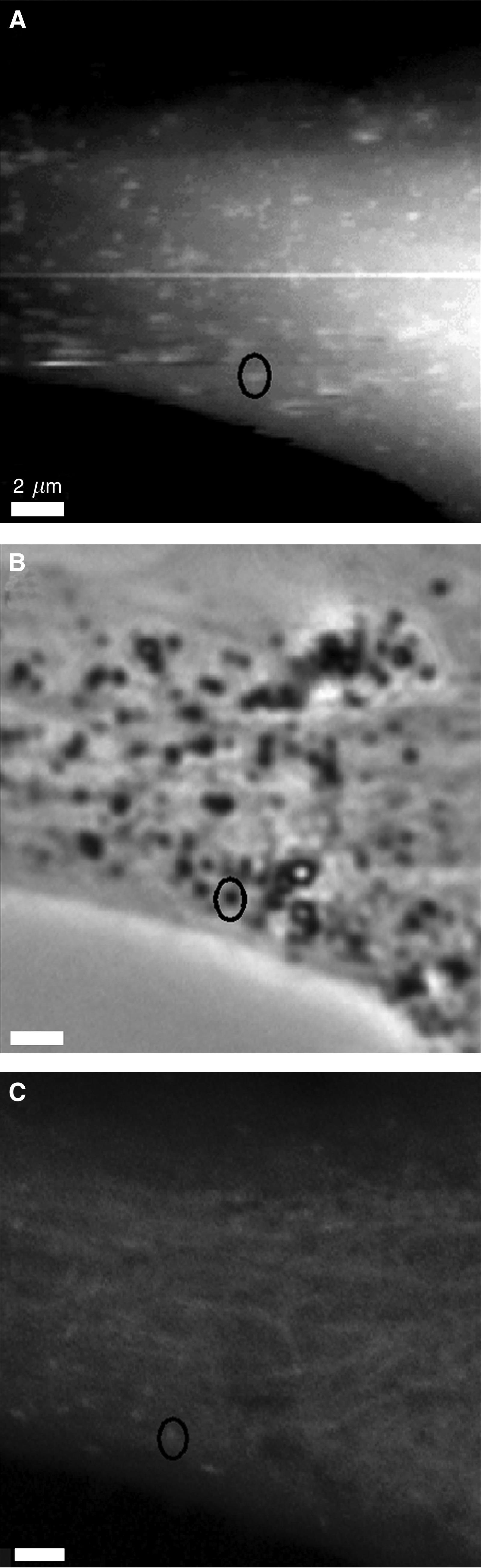
The protrusions on the surface of melanocytes correspond to actin-associated vesicles. The AFM topograph (A; height range: 4.3 μm) and corresponding phase-contrast image (B) of melanocytes treated with FITC-phalloidin show that some of the protrusions observed in the AFM topograph correspond to vesicles. Additionally, the LSCM image (C; optical slice: 0.8 μm) shows brighter actin staining at a fraction of the corresponding points. The AFM image was recorded in the trace scan direction.