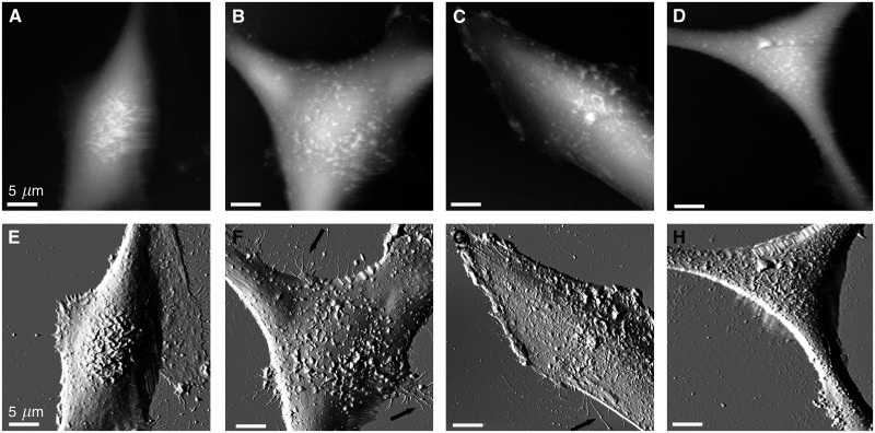
Figure 3.
Melanoma cell surfaces from VGP tumours are characterised by highly flexible ridges. The AFM topographs (A–D) and corresponding error signal images (E–H) reveal flexible ridges on the surface of four distinct melanoma cell lines, SK-Mel-28 (A, E), WM-115 (B, E), WM-853-2 (C, G) and WM-39 (D, H). Arrows indicate filopodial extensions. Height ranges correspond to (A) 8.7 μm, (B) 8.4 μm, (C) 6.9 μm and (D) 7.8 μm. Images presented were recorded in the trace scan direction.

