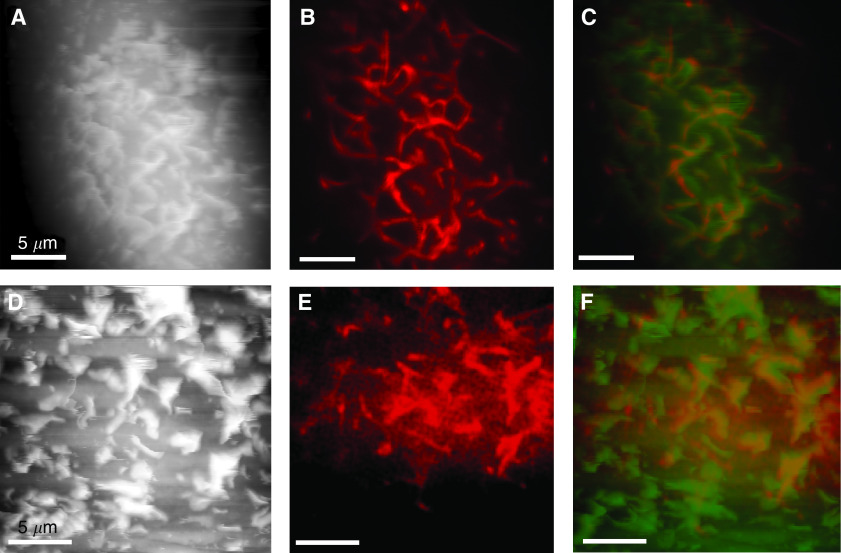
Figure 4.
Flexible structures on the surface of VGP melanoma cells are actin based. The AFM topographs (height ranges: (A) 6.7 μm; (D) 6.7 μm) and LSCM images (B, E; optical slice: 0.8 μm) of cells treated with TRITC-labelled phalloidin reveal that the flexible structures on the surface of VGP melanoma cells are actin based, in cell lines SK-Mel-28 (A–C) and WM-115 (D–F). Overlay images (C, F), with the AFM topograph in false green and confocal images in red, highlight the correlation. The AFM topographs presented were recorded in the trace scan direction.

