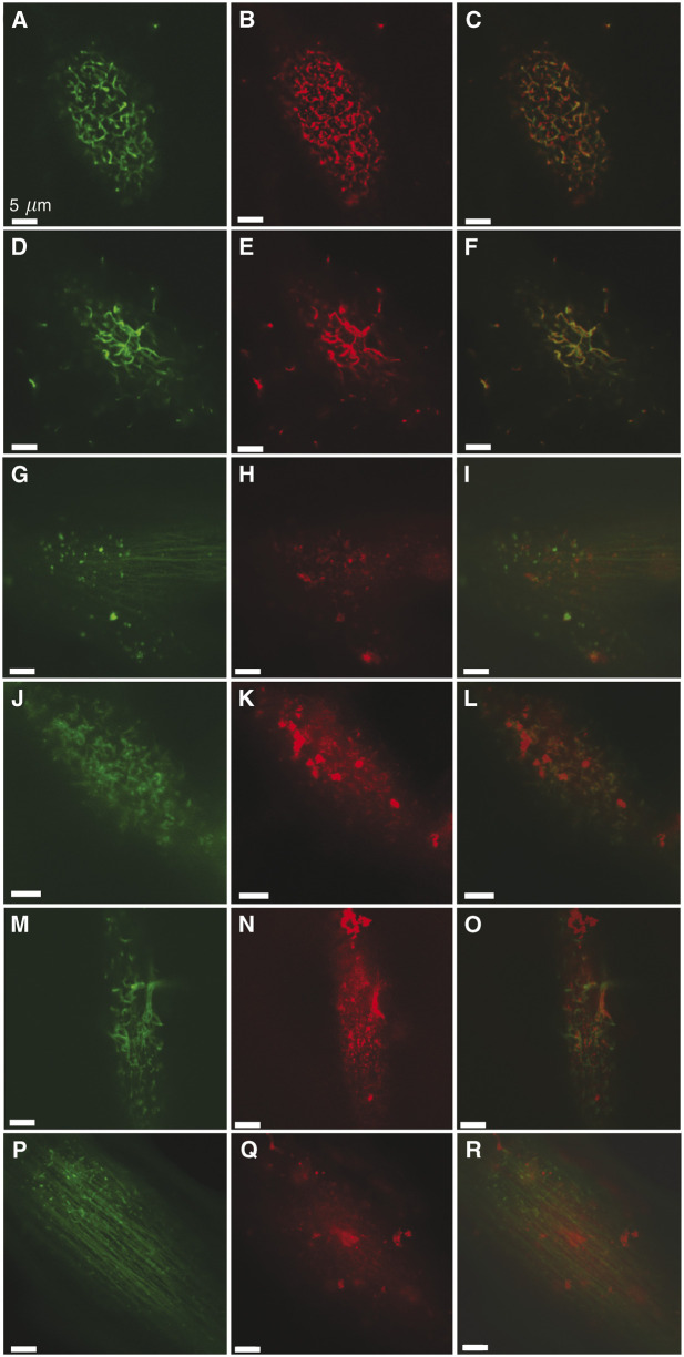
Figure 5.
The β1 integrin colocalises with subsurface actin structures. The LSCM images (optical slice: 0.8 μm) of SK-Mel-28 (A–C) and WM-115 (D–F) cells, dual labelled with FITC-phalloidin (A, D) and anti-β1 integrin antibody, detected using TRITC-conjugated goat anti-mouse secondary antibody (B, E), reveal that the β1 integrin distribution correlates with the subsurface actin structures (overlays: C, F). The β1 integrin was more diffusely distributed on the surface of melanocytes, with some association with actin-associated protrusions noted (labelled actin (G), labelled fibronectin (H), overlay (I)). The LSCM images of SK-Mel-28 (J–L) and WM-115 (M–O) cells dual labelled with FITC-phalloidin (J, M) and anti-fibronectin antibody detected using TRITC-conjugated secondary antibody (K, N) show that fibronectin is partially bound at the actin-based ridges (overlays: L, O). In comparison, the LSCM images of melanocytes (P–R) dual labeled with FITC-phalloidin (P) and anti-fibronectin detected using TRITC conjugated antibody (Q) do not reveal a correlation between actin-based surface structures and bound fibronectic (overlay, R).

