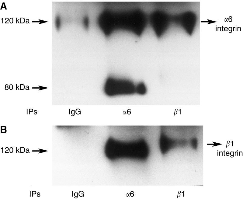
Figure 2.
α6β1 interaction in HEY cells. Monolayer cultures of HEY cancer cell line was washed twice with PBS and harvested with trypsin-versene. Cells were lysed in lysis buffer (100 mM Tris-HCl, pH 7.5, 150 mM NaCl, 1 mM CaCl2, 1% Triton X-100, 0.1% SDS, 0.1% NP-40, 1 mM vanadate, 1 μg ml−1 pepstatin, 1 mM PMSF, 5 μg ml−1 Trasylol and I μg ml−1 of leupeptin). Protein concentrations of the cell lysates were determined and lysates containing equal protein were used for immunoprecipitation. Cells lysates were immunoprecipitated with mAbs against against α6 (43B-9B), β1 (PD52) or isotype matched control. Samples were resolved in 7.5% SDS–PAGE gel under nonreducing conditions and transferred to nitrocellulose membranes. Membranes were then probed with (A) anti-α6 integrin; (B) anti-β1 integrin antibodies and the interaction of α6β1 integrin was evaluated with ECL.

