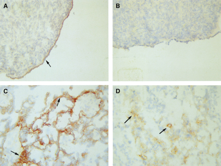
Figure 3.
Expression of α6 integrin and uPAR in normal ovaries and ovarian tumours. Cryostat sections of ovarian tissues were stained by the immunoperoxidase method for the expression of α6 integrin and uPAR as discussed in the Materials and Methods section. (A) Normal ovary, arrow showing continuous basal expression of α6 integrin in the epithelium; (B) normal ovary, showing no expression of uPAR; (C) grade 3 serous ovarian tumour, arrows indicating irregular expression of α6 integrin in the basal epithelial and (D) grade 3 serous ovarian tumour, arrows indicating cytoplasmic epithelial staining of uPAR.

