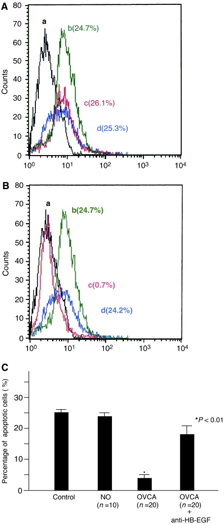
Figure 2.
Cell survival activity mediated by peritoneal fluid in patients with a normal ovary or ovarian cancer. Flow cytometric analysis for apoptotic cells in SKOV3 cells after incubation with peritoneal fluid of a normal ovary (A) and ovarian cancer (B). Control (a: black line). Under serum-free condition (b: green line). Incubation with 10% peritoneal fluid in the absence (c: red line) or presence (d: blue line) of an inhibitory antibody against HB-EGF. Each percentage indicates the ratio of apoptotic cells in SKOV3 cells. (C) Alteration in the percentage of apoptotic cells after incubation with the peritoneal fluid from a normal ovary or ovarian cancer. A bar indicates the mean value and standard errors. The P-value represents comparison with the levels of patients with a normal ovary.

