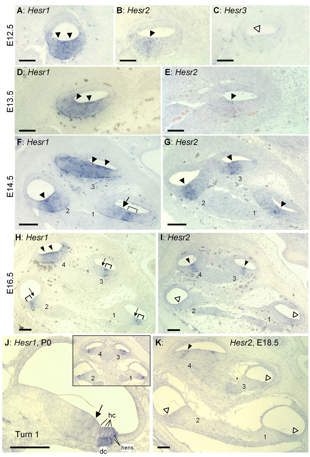
Fig. 1. Expression patterns of Hesr1 and Hesr2 in the developing cochlea.

A–E: Adjacent sections of the middle part of the E12.5 (A–C) and E13.5 (D,E) cochlear duct. Expression of Hesr1 or Hesr2 are indicated by solid arrowheads. No expression in the prosensory domain was detected by Hesr3 probes at this stage (open arrowhead in C). Adjacent sections of E14.5 (F, G), E16.5 (H, I) and E18.5 (K) cochlea. Levels (half-turns) of cochlear duct are numbered from base (turn 1) to apex (turn 3 or turn 4). Open arrowheads in I and K indicate no expression. J: Higher magnification micrograph of the basal turn (turn1) of P0 cochlea. Inset shows entire cochlea. Arrowheads, arrow and brackets indicate expression domains. dc: Deiters’ cells. hc: Hair cells. hens: Hensen's cells. Scale bar= 100 µm.
