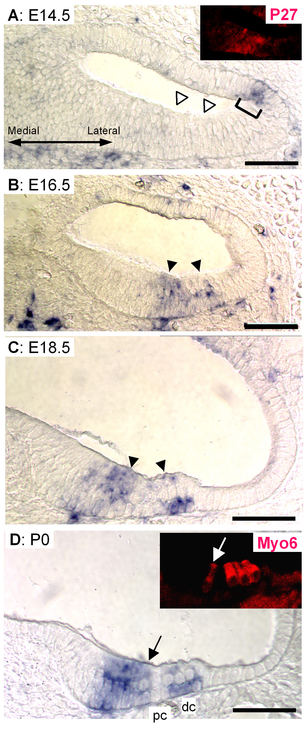
Fig. 2. Expression patterns of Hesr3 in the development of cochlea.

E14.5 cochlea (A) did not show Hesr3 signal in the developing sensory epithelium (white arrowheads); however, lateral side of cochlea duct showed some expression (bracket). Inset in A shows immunostaining of the same section with anti-p27kip1. E16.5 (B) and E18.5 (C) samples clearly showed the expression of Hesr3 in the sensory epithelium (black arrowheads in B, C). D shows P0 cochlea. Hesr3 mRNA was detected in the sensory epithelium and GER. Inset in D shows immunostaining of the same section with anti-Myo6. Arrow indicates inner hair cell. dc: Deiters’ cells. pc: Pillar cells. Scale bar= 50 µm.
