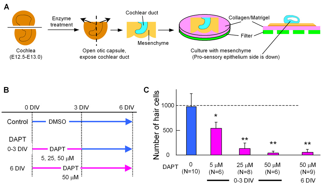
Fig. 6. DAPT treatment inhibits hair cell differentiation.

Schematic of culture method and experimental design of DAPT treatments (A, B). E12.5–13.0 cochlear ducts were cultured with surrounding mesenchyme on collagen/Matrigel (A). The cochleas were separated into control and incubated with 3 different concentrations of DAPT for 2 different durations (B), and hair cells were counted after 6 days in vitro (DIV). C: Number of hair cells in a cochlea cultured for 6 days with or without DAPT according the time schedule in A. Error bars indicate standard deviations of the means. N= number of cochleas. Asterisk indicates P< 0.05, double asterisk indicate P< 0.005 compared to the control with a Student's T-test.
