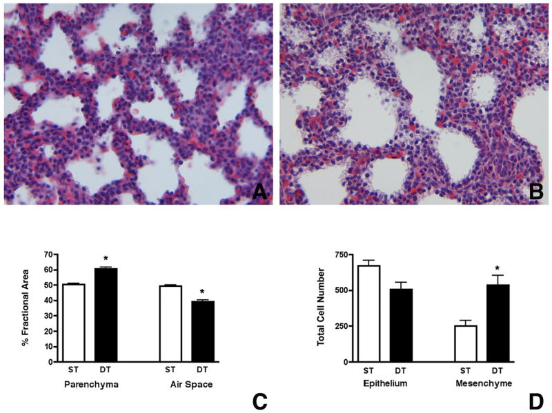
Figure 7.

Misexpression of Mia1 results in mesenchymal thickening and reduced alveolar space. Mia1 expression was induced in the epithelium of SP-C/MIA mice beginning on E16.5 and continued until sacrifice at E18.5. Histology shows that the lung mesenchyme in SP-C/MIA double transgenic fetuses (B) appears thickened compared to single transgenic (expressing only the tetO/MIA transgene) littermates (A). Comparison of changes in fractional areas of airspace and respiratory parenchyma in the E18.5 lungs show that SP-C/MIA double transgenic (DT) mice have significantly (*, p < 0.001) more parenchyma and less airspace compared to single transgenic (ST) mice (C). Comparison of total cell number show that total mesenchymal cells are significantly (*, p < 0.01) higher in SP-C/MIA mice compared to controls while no significant differences are observed in total epithelial cells between the two groups (D). Data are plotted as means ± SE (n = 4).
