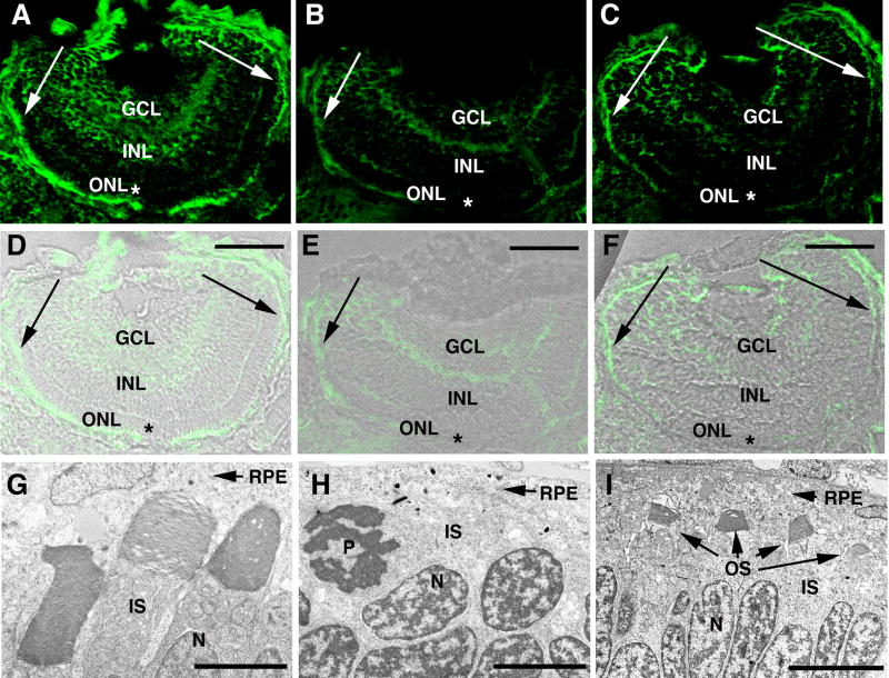
Figure 6. Correlation between photoreceptor OS loss and extent of Kif17 knockdown in morphants.
A–C Kif17 distribution in eyes of 3 day old control (A) and morphant larvae (B,C). D–F. IC images A–C merged with bright-field images of the same sections. Abbreviations: GCL, ganglion cell layer; INL, inner nuclear layer; and ONL, outer nuclear layer. Scale bars in D–F = 40 μm. Note that in the photoreceptor layer of the early developing central retina ( asterisks) of morphants Kif17 immunoreactivity is almost completely lacking but in the late developing periphery (indicated by arrows), Kif17 protein is present. G–I. EM images from the central region of a control retina (G), and of a morphant retina (H). I is from the peripheral region of a morphant retina. Note that OS form throughout the retina of controls and are virtually absent from the central retina of morphants where Kif17 protein knock-down is most complete. OS are seen in the periphery of some but not all morphants. EM magnifications bars: 2.8 μm (G), 4.0 μm (H), and 6.8 μm (I).

