
Fig. 2.
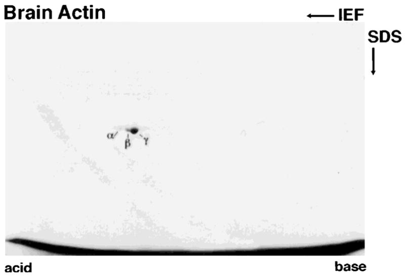
Two-dimensional gel of brain actin stained with Coomassie Blue. A major spot corresponding in pI to γ-actin (pI 5.3) and two minor spots corresponding in pI to β-actin (pI 5.28) and smooth muscle α-actin (pI 5.2) are resolved.
Official websites use .gov
A
.gov website belongs to an official
government organization in the United States.
Secure .gov websites use HTTPS
A lock (
) or https:// means you've safely
connected to the .gov website. Share sensitive
information only on official, secure websites.
Two-dimensional gel of brain actin stained with Coomassie Blue. A major spot corresponding in pI to γ-actin (pI 5.3) and two minor spots corresponding in pI to β-actin (pI 5.28) and smooth muscle α-actin (pI 5.2) are resolved.