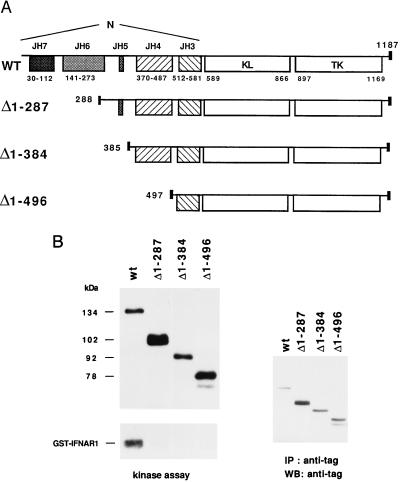
Figure 1.
(A) Schematic representation of wt and deleted Tyk2 proteins. The position and size of the JH domains and the first amino acid of each mutant protein are indicated. The small black box corresponds to the VSV-G epitope. (B) Basal in vitro kinase activity of wt and deleted Tyk2 forms. Whole cell extracts of the indicated transfectant were immunoprecipitated with anti-VSV-G antibodies. One-third of the immunoprecipitate was subjected to an in vitro kinase assay (Left) and the remaining material was directly resuspended in SDS/sample buffer. Samples were then fractionated on SDS/7% polyacrylamide gel, transferred to Hybond C-super membrane and subjected to autoradiography (Left) or probed with a monoclonal anti-VSV-G antibody, followed by goat anti-mouse 125I-conjugated, and subjected to autoradiography (Right). Phosphorylated and iodinated bands were quantified with a PhosphorImager.

