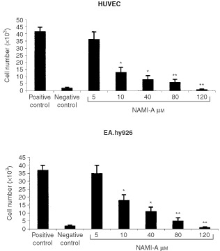
Figure 1.

Effect of NAMI-A upon endothelial cell proliferation. Low density cultures of endothelial cells (2.5×10³ well−1) were exposed on day 0, 2 and 4 with complete medium (positive control), serum-free medium (negative control) or complete medium containing different NAMI-A concentrations. Cell number was evaluated on day 6 by Kueng et al's method (1989). Bars, means±s.d. of three independent experiments. *P<0.05 and **P<0.01, Student–Newman–Keuls analysis of variance.
