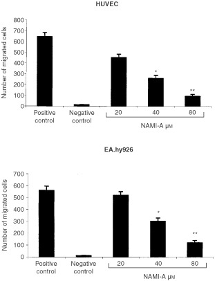
Figure 2.

Effect of NAMI-A upon endothelial cell chemotaxis. 1.2×10⁵ cells, exposed for 24 h to each dose of NAMI-A, were seeded in the upper compartment of the Boyden chamber, and the conditioned medium of NIH3T3 cells was placed in the lower compartment as the chemoattractant. Unexposed cells were used in the positive and negative control, respectively, the latter being devoid of the chemoattractant. Cells that migrated after 6 h incubation to the lower surface of the filter separating the compartments were counted. Bars, means±s.d. of the number of migrated cells in five to eight 400x fields of three filters per specimen. *P<0.05 and **P<0.01, Student–Newman–Keuls analysis of variance.
