
Figure 4.
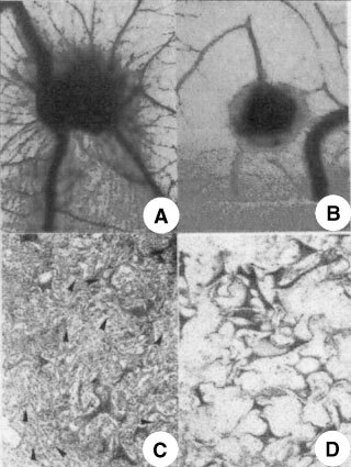
The CAM of a 12-day-old chick embryo incubated for 4 days with a gelatin sponge loaded with (A), (C) the angiogenic fibroblast growth factor-2 (FGF-2) or with (B), (D) 240 μM of NAMI-A. Note in (A) numerous blood vessels converging like spokes toward the sponge, whereas in (B) there are very few vessels around the sponge or converging toward it. (C) Histologic section of the sponge shows numerous vessels (arrows) intermingled in a collagenous matrix among the trabeculae. (D) No vessels are detectable. Original magnifications: A, B, 50×; C, D, 400×.