Figure 1.

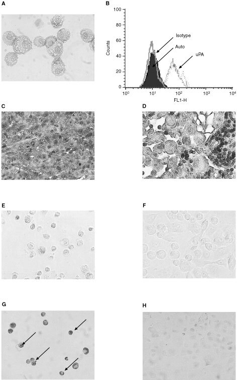
uPA expression in PC3 cells, PC3 xenografts and metastatic lymph nodes and TUNEL assay of PC3 cells in vitro. (A) PC3 cells are strongly positive to membrane-bound uPA. (B) Flow cytometry histogram of PC3 cells showing indirect immunofluorescence labelling of cell surface uPA. Auto: autofluorescence level; Isotype: mouse anti-human subclasses IgG1 control antibody; uPA: mouse anti-human uPA IgG1 antibody. (C) PC3 tumour xenograft cancer cells are strongly positive to uPA. (D) Cancer cells from metastatic lymph nodes are positive to uPA while lymphocytes are negative to uPA (small cells). The sections were from different mice. PC3 cells were either treated with 213Bi-PAI 2 (E) or untreated (F). After 36 h, the treated and untreated cells were fixed and processed for TUNEL assay. The cells were then visualised by light microscope. The arrows represent typical apoptotic cells with condensed or fragmented nuclei (G), while control cells show normal shapes (H). A, C, E, F, G and H: Magnification ∼×200. D: Magnification ∼×400.
