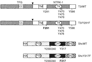
Figure 1.

Schematic representation of TRK-T3 and Shc constructs. In the TRK-T3 constructs the portions contributed by TFG and NTRK1 are shown, with an arrowhead indicating the breakpoint. The coiled-coil (CC), transmembrane (TM) and tyrosine kinase (TK) domains are indicated. The tyrosine residues involved in Shc and FRS2 interaction (Y291), PLCγ interaction (Y586) and tyrosines of the activation loop (Y470, Y475 and Y476) are indicated. In T3/Y291F mutant the tyrosine 291 has been mutated to phenylalanine (F291). The TRK-T3 cDNAs were inserted into the pRC/CMV expression vector. The Shc constructs show the PTB domain, the collagen homology region (CH1) and the SH2 domain. Y239/240 and Y317 are tyrosine residues phosphorylated by tyrosine kinases. In ShcY317F, tyrosine 317 is mutated to phenylalanine (F317). The Shc cDNAs contain the HA epitope at the N-terminus and were inserted into the pCGN mammalian expression vector.
