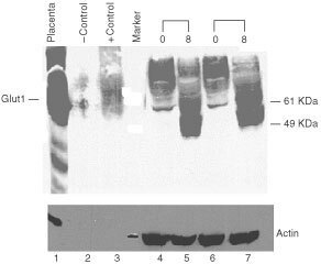
Figure 7.

Glut-1 transporter protein levels in breast cancer cells lines treated with 2-DG. SkBr3 breast cancer cells were treated with 2DG for 48 or 72 h with 8 mM 2DG (8) or without 2DG (0). Protein was isolated, size separated on a 10% polyacrylamide gel. A Western blot was performed using a polyclonal anti-Glut-1 antibody. Positive controls are protein isolated from human placenta and membranes isolated from Glut1 injected Xenopus oocytes (+control). Sham injected oocytes were used as a negative control (−control). Glut1 protein is indicated. Molecular weight markers are labelled. The protein blot was reacted with anti-actin antibody after removal of the Glut1 antibody (lower panel).
