
Figure 4. Misrouting of Odorant Receptor-Specific Olfactory Axons in the Absence of IGF Signaling.
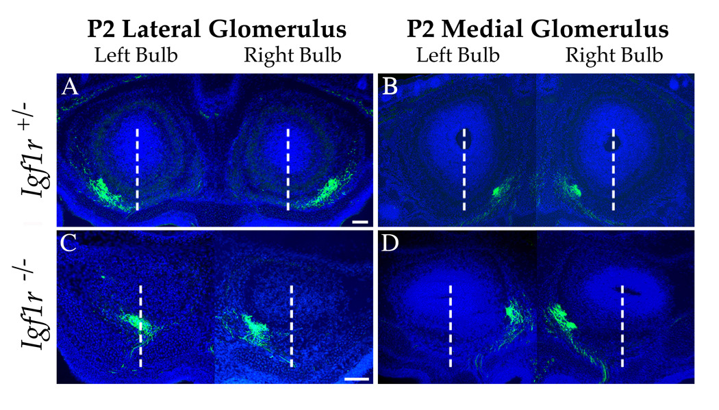
To localize the glomeruli of one class of odorant-specific olfactory axons, E18.5 P2-IRES-tau:lacZ mice either heterozygous (A and B) or nullizygous (C and D) at the Igf1r locus were stained with an anti-β-galactosidase antibody (green). (A, B) In Igf1r+/− mice, P2 neurons project to two glomeruli per olfactory bulb, one on the lateral (A) and, more caudally, one on the medial (B) face of the bulb. The position of these glomeruli is stereotyped between animals. (C, D) P2 neurons still form two glomeruli in the Igf1r nullizygous background. However, the lateral glomerulus (C) is shifted toward the medial hemisphere of the bulb, resulting in a distortion of the medial-lateral mirror symmetry of olfactory bulb innervation. Dashed white lines demarcate the midlines of each bulb. Note that in this particular individual, the lateral glomerulus in the right-hand bulb has shifted well beyond the midline. The medial glomeruli in the Igf1r nullizygote (D) appears to be in a position similar to that observed in the control (B), with only a small dorsomedial shift, which is likely due to the compression of the map into a smaller area of the bulb’s surface. In this example, the medial P2 axons form a doublet in the right bulb (D), a common phenomenon seen with this glomerulus (Royal and Key, 1999). Images of the left and right bulb are combined for clarity. Nuclei were stained with Hoechst 33342 (blue). Scale bar = 100µm.