
Figure 6. Fluorescent immunomicroscopy of IGFBP-3 and DAPI Stained Nuclei in Young Human Cartilage.
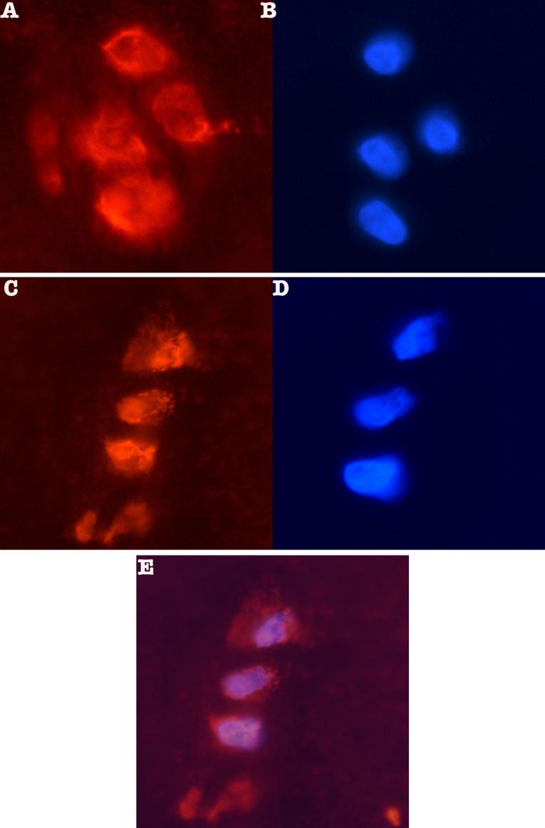
Panels A and C show the orange fluorescent stain for IGFBP-3 detected with Upstate anti-IGFBP-3 (1:250 dilution) and a CY-2 conjugated goat anti-rabbit secondary antibody (Jackson Immunoreasearch). Panels B and D show adjacent sections stained with DAPI to reveal the nuclei. Panel E shows an overlay of panels C & D demonstrating co-localization of IGFBP-3 and DAPI stain.