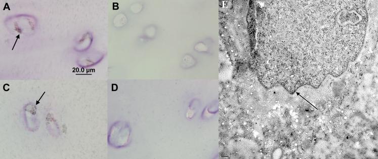
Figure 7. Immunogold and Immunohistochemical Staining on Thin Sections of Human Cartilage.
Panels A through D show light micrographs of 1 μm sections. Panels A and C show staining of nuclei in chondrocytes (arrows) in the transitional zone and in the superficial zone respectively. Panels B and D show negative controls (without antibody). Panel F shows intra-nuclear immunogold labeling (arrow) for IGFBP-3 in a transitional zone chondrocyte imaged by TEM.

