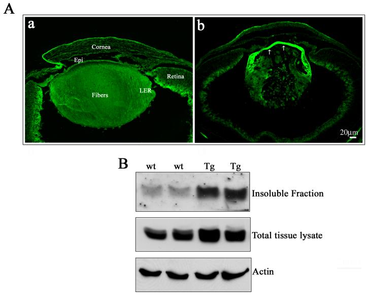
Fig. 9. Increased αB-crystallin phosphorylation in the RhoGDIα transgenic lens epithelium.
A. P1 WT and Tg lens cryosections immunostainined with a Ser-59 phosphospecific αB-crystallin antibody revealed the presence of phosphorylated αB-crystallin in the epithelium and fibers cells (a). However, while distribution of phosphorylated αB-crystallin was uniform between the epithelium and fiber cells of WT lenses, the Tg lenses (b), exhibited a very intense staining for phosphorylated αB-crystallin throughout the epithelium, including at the equatorial and central regions (arrows). A representative photograph is presented in this figure based on multiple analyses using lens sections derived from the different Tg lines. B. Transgenic lens total (800x supernatants) or insoluble fractions (100,000xg pellets) immunoblotted with phosphospecific αB-crystallin antibody showed a significant increase in the levels of phosphorylated αB-crystallin, both in total homogenate and membrane fractions compared to the WT lenses. Actin was probed in the same samples to confirm loading equivalence for protein.

