Figure 1.
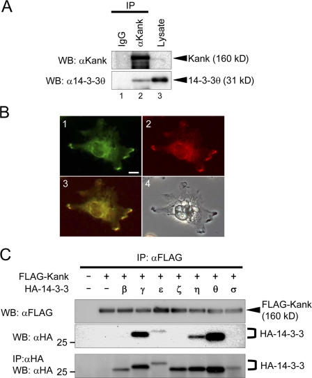
Kank interacts with 14-3-3 isoforms γ, ε, η, and θ. (A) Kank interacts with 14-3-3θ. VMRC-RCW cells were lysed and the lysate was immnunoprecipitated (IP) with control rabbit IgG (lane 1) or anti-Kank pAb (lane 2). The immunoprecipitates and the lysate (lane 3) were subjected to Western blotting (WB) using the antibodies indicated. (B) Endogenous Kank and 14-3-3 proteins are colocalized in VMRC-RCW cells. A fluorescence image of Kank (green, lane 1), a fluorescence image of 14-3-3 (red, lane 2), a merged image of lanes 1 and 2 (yellow, lane 3), and a light microscope image (lane 4) are shown. Bar, 10 μm. (C) Kank associates with 14-3-3 isoforms γ, ε, η, and θ. HEK293T cells were transfected with the indicated expression vectors. Immnunoprecipitation was performed using an anti-FLAG antibody and the bound proteins were detected by Western blotting using the antibodies indicated.

