Figure 5.
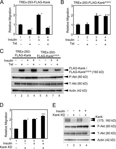
Kank inhibits cell migration through 14-3-3 and partially inhibits insulin-mediated cell migration. (A and B) Tetracycline-inducible TREx-293 cells stably expressing Kank (A) or KankS167A (B) were subjected to cell migration assays using TransWell. TREx-293 cells expressing tetracycline-inducible FLAG-Kank or FLAG-KankS167A with (lanes 3 and 4) or without (lanes 1 and 2) insulin (10 μg/ml) stimulation were used for cell migration assays. The ratios of migration compared with the control were calculated. The results shown are the means ± SD of triplicate experiments. Tet, tetracycline induction. *, P < 0.01 compared with the control ; **, P < 0.05 compared with the control. (C) The expression of FLAG-Kank and FLAG-KankS167A is induced in tetracycline-inducible TREx-293 cells. The expression of FLAG-Kank (lanes 1–4) and FLAG-KankS167A (lanes 5–8) with or without the treatment with 10 μg/ml insulin and/or tetracycline was examined by Western blotting. (D) Knockdown of Kank enhances cell migration. The esiRNA of Kank (Kank KD, lanes 2 and 4) and control esiRNA (lanes 1 and 3) were prepared as described in Materials and methods and transfected into HEK293 cells. Cell migration assays were performed using these transfected cells in the presence (lanes 3 and 4) or absence (lanes 1 and 2) of 10 μg/ml insulin. The relative migration was calculated as described in A. *, P < 0.01 compared with the control; ***, P < 0.01 compared with the control and P < 0.05 compared with Kank KD. (E) The expression of endogenous Kank is decreased by the esiRNA of Kank. The expression levels of endogenous Kank were determined by Western blotting. The cells were transfected and stimulated as described in D.

