
Figure 1.
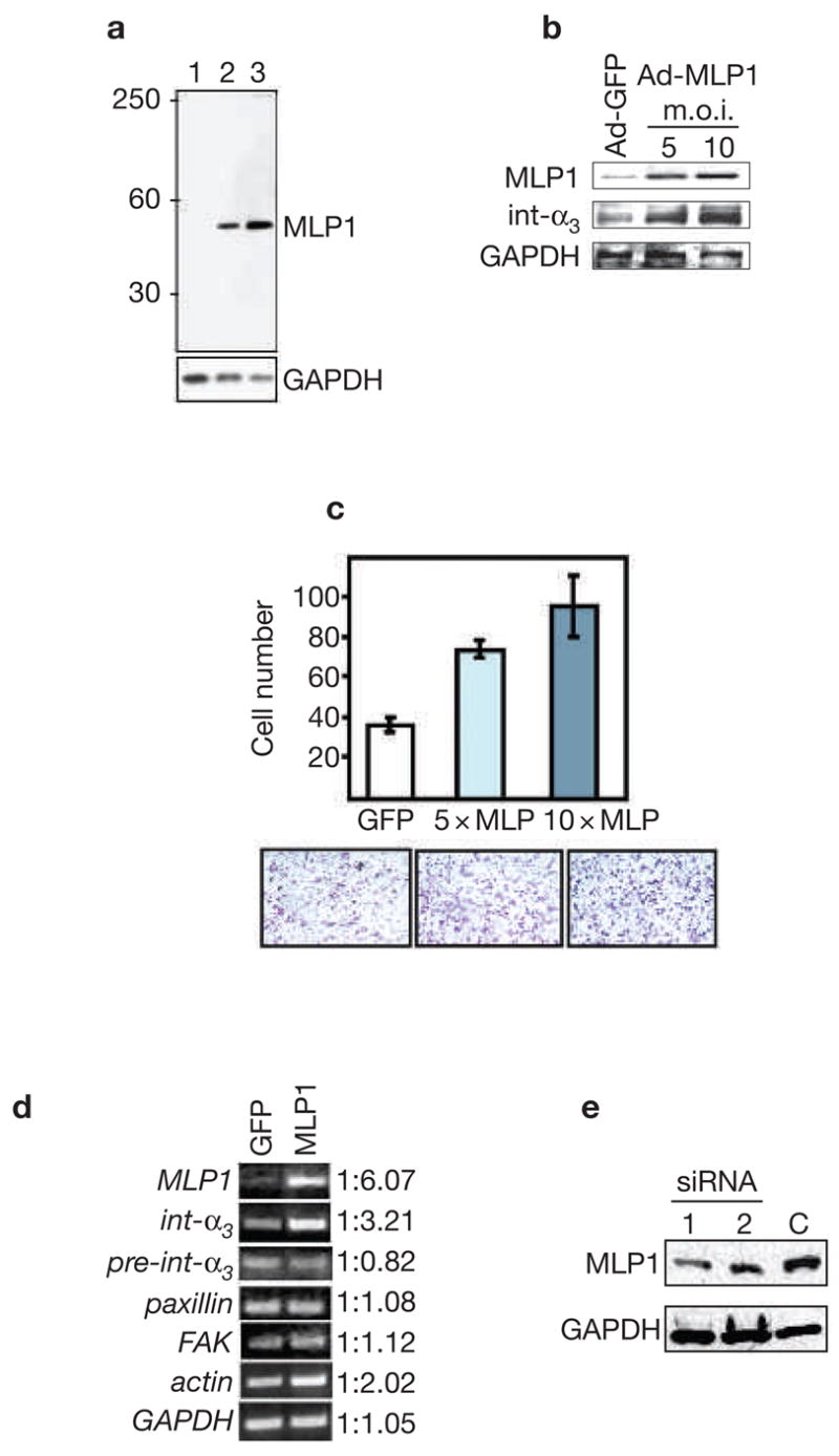
MLP1 regulates integrin α3 expression at the post-transcriptional level. (a) Protein extracts (10 μg/lane) from three cell lines were probed with rabbit anti-MLP1 antibody. The blot was stripped and re-probed for glyceraldehyde-3-phosphate dehydrogenase (GAPDH) as the loading control. Lane 1: H520 cells; lane 2: A549 cells; lane 3: HT29 cells. A single band of expected size (relative molecular mass of approximately 40,000) was detected. MLP1 was not detectable in H520 cells with the amount of total protein extracts used. (b) H520 cells were infected with empty adenoviral vector (Ad-GFP; m.o.i. 10) or vector containing the MLP1 coding sequence (Ad-MLP1) at m.o.i. of 5 or 10. Total protein extracts (50 μg; higher amount needed for detecting integrins) were probed with the antibodies indicated on the left. GAPDH served as the loading control. Increased expression of MLP1 correlates with increased expression of integrin α3. (c) H520 cells were transfected with adenoviral vector expressing green fluorescent protein (GFP) alone (at 10× m.o.i.), or GFP plus MLP1 (at 5× and 10× m.o.i.), and plated onto the upper chamber of the transwell. Cells that migrated to the underside of the upper chamber were stained with crystal violet and the numbers compared. Lower panels show representative views of the migrated cells. (d) Reverse transcription polymerase chain reaction (RT-PCR) analysis using primers specific for MLP1 mRNA, integrin α3 exon (int-α3), integrin α3 intron–exon junction (pre-int-α3), or mRNAs of paxillin, focal adhesion kinase (FAK), actin and GAPDH. Total RNAs were isolated from H520 cells infected with adenoviral vector (GFP) or vector containing the MLP1 coding sequence at 5× m.o.i. The linear range of PCR amplification was empirically determined for each primer set (see Methods). At the mid-point of the linear curve, amplified samples were run on ethidium-bromide-stained agarose gel and quantified. Integrin α3 RNA level increases in step with increased expression of MLP1, whereas the pre-spliced integrin α3 RNA level remains unchanged. There is a moderate increase of actin in the presence of MLP1 and no change for paxillin, FAK or GAPDH. Note that without reverse transcriptase, the PCR reactions did not yield any amplified products (data not shown). (e) A549 cells were transfected with empty vector (C) or vector containing one of the two MLP1-specific small interfering RNA (siRNA)-encoding oligonucleotide duplexes.