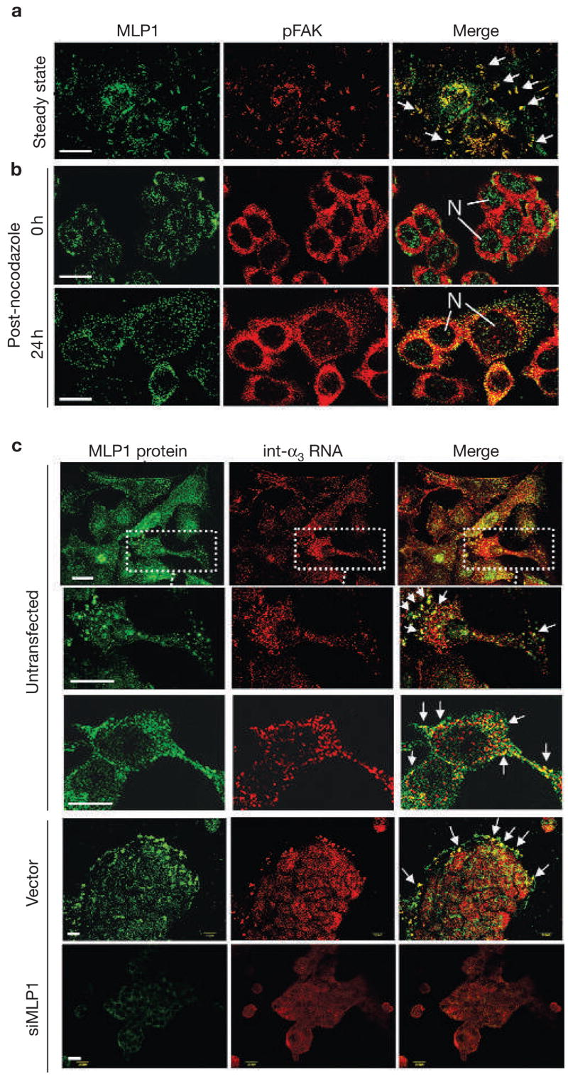
Figure 3.

MLP1 regulates localization of integrin α3 RNA. (a) A549 cells were double-stained for MLP1 (green) and phospho-focal adhesion kinase (pFAK; red). Arrows mark examples of prominent adhesion plaques, where MLP1 and pFAK colocalize. (b) HT29 cells were treated with nocodazole and then returned to normal media. Cells were fixed and double-stained for MLP1 (green) and pFAK (red) at 0 and 24 h after recovery. Before recovery from nocodazole treatment (0 h), MLP1 was mainly localized in the nuclei (N). Twenty-four hours after recovery, MLP1 distribution returned to a punctated pattern with substantial colocalization with pFAK. (c) HT29 cells were double-stained for MLP1 protein and integrin α3 (int-α3) RNA. Untransfected cells and cells transfected with empty vector or vector expressing MLP1 small interfering RNA (siRNA) are indicated. In untransfected samples, a cell cluster and two enlarged single-cell views are shown to better visualize the large complexes that have been co-stained for integrin α3 RNA and MLP1 protein. In vector and siMLP1-transfected samples, clusters of cells are shown and the integrin α3 RNA–MLP1-containing complexes are mostly at the leading edges (arrows). Sense integrin α3 RNA probe showed no signals (data not shown). Scale bars, 20 μm.
