Figure 5.

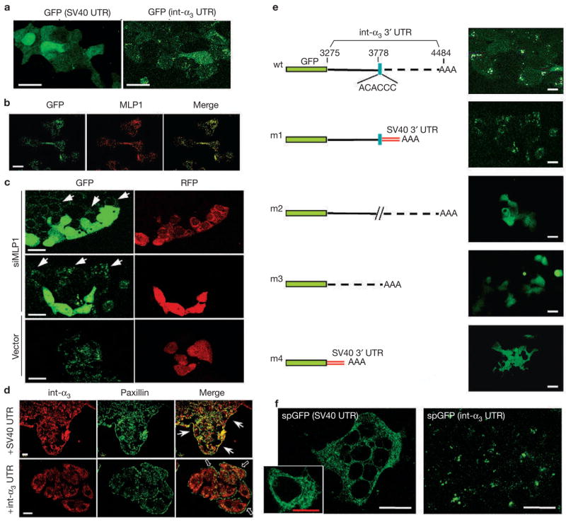
Localized GFP expression mediated by the zipcode in integrin α3 3′ UTR. (a) HT29 cells were transfected with vectors containing enhanced green fluorescent protein (eGFP) open reading frame upstream of the SV40 3′ untranslated region (UTR) or the integrin α3 (int-α3) 3′UTR, and observed for GFP expression. With SV40 3′ UTR, GFP showed a diffuse expression pattern throughout the cell body. With integrin α3 3′ UTR, GFP was localized in a punctated pattern. (b) The same HT29 cells that were transfected with GFP–integrin-α3 chimaeras were fixed and stained for GFP and MLP1. The two proteins colocalized in subcellular punctates. (c) HT29 cells expressing GFP–integrin-α3 chimaeras were transfected with vector containing red fluorescent protein (RFP) alone (vector) or RFP and MLP1 small interfering RNA (siRNA) #1 (siMLP1; see Fig. 1). Live cells were examined. In the presence of siMLP1 (co-expressing RFP) the GFP became diffuse, whereas in neighbouring untransfected cells (arrows) the GFP remained in a punctated pattern. Transfection by the vector alone did not have any effects on the punctated GFP expression. (d) Transfected HT29 cells (+SV40 UTR or +int-α3 UTR) were stained for the endogenous integrin α3 (red) and paxillin (green). Ectopic overexpression of integrin α3 3′ UTR resulted in dispersion of endogenous integrin α3 protein from the paxillin-containing plaques (empty arrows), whereas overexpression of SV40 3′ UTR had little effect. Arrows point to plaques containing integrin α3 and paxillin at the leading edges. (e) Various deletion constructs of the integrin α3 3′ UTR were tested for the essential cis-acting localization element. AAA depicts the polyadenylation signal. SV40 3′ UTR that was contained in the cloning vector provided the default polyadenylation signal for the m1 and m4 constructs. The zipcode (blue box) seemed to be the most critical cis element. wt, wild type. (f) Chimeric integrin α3 signal peptide (sp)-GFP–SV40 or spGFP–integrin-α3 3′ UTR were transfected into HT29 cells and the GFP patterns were observed. With SV40 3′ UTR, the expected membrane-targeted GFP (spGFP) appears associated with an intracellular network, presumably the ER. The inset shows an enlarged view of a single cell displaying the network-bound GFP. With the integrin α3 3′ UTR, the GFP remains in the same punctated pattern as without the signal peptide. Scale bars, 20 μm, except the red bar in the inset, which is 10 μm.
