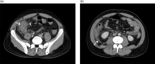
Figure 5.
(a) Axial MDCT image of a patient presenting with typhlitis following chemotherapy. Note the caecal thickening with mucosal enhancement (arrow) and associated peri-colic mesenteric stranding (arrowhead). (b) Axial MDCT image of a neutropenic patient with typhlitis showing a thickened, enhancing ascending colon, peri-colic fluid (arrow) and mesenteric stranding. Note the relative sparing of the transverse colon and proximal small bowel loops.

