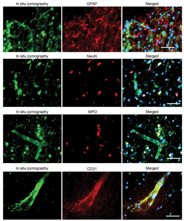
FIG. 7.

Representative photomicrographs of double fluorescent staining of in situ zymography and different cell markers. In situ gelatinolytic activity was seen as green fluorescence. GFAP, NeuN, MPO, and CD31 were seen as red fluorescence. Merged photos showed that GFAP staining did not colocalize with in situ gelatinolytic activity. Some NeuN staining colocalized with in situ gelatinolytic activity. Most of the MPO staining colocalized with in situ gelatinolytic activity. CD31 staining colocalized with in situ gelatinolytic activity on the vessel wall. Scale bar = 50 μm.
