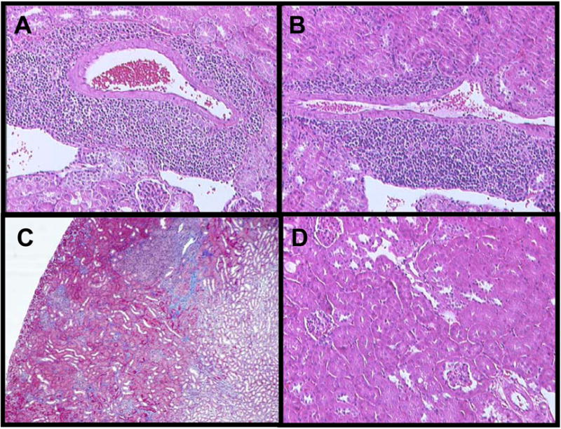
Figure 4. Histologic features in accepted renal allografts.

Renal grafts from C57BL/6 recipients were collected and formalin fixed sections were prepared and stained with H&E or trichrome. Magnification X100. Prominent perivascular mononuclear cell infiltration is observed in accepted renal allografts collected at 60 days post-transplant (A) and at 150 days post-transplant (B). Mild interstitial fibrosis is observed in renal allografts at 150 days post-transplant (C). The renal parenchyma of accepted renal isografts at day 150 post-transplant is normal (D).
