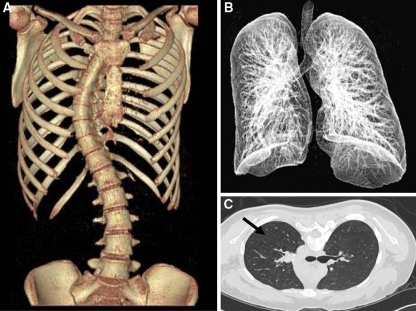
Fig. 2.
The individual right and left lung volumes were measured in each case using the 3D image reconstruction software (RAPIDIA 2.7, INFINITT, South Korea). This program recognizes, the “Air density shade” of the lung (shown here with arrow), and volume for every section of the lung; which then automatically calculate the volume for individual lung by summation of all section volumes. a 3D CT scan image showing scoliosis of the spine with the rib cage. b 3D CT scan image of lung fields. c Cross sectional CT scan image used to calculate lung volume

