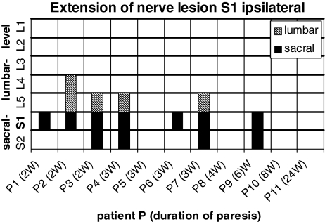
Fig. 2.
Distribution of pathological spontaneous activity on the ipsilateral side in 11 patients with a S1 nerve root lesion in cranial and caudal extension. The filled bars correspond to axonal lesion signs in the sacral segments, the hatched bars to axonals lesion signs in the lumbar segments of each patient. Duration of the paresis in weeks (w)

