Fig. 1.

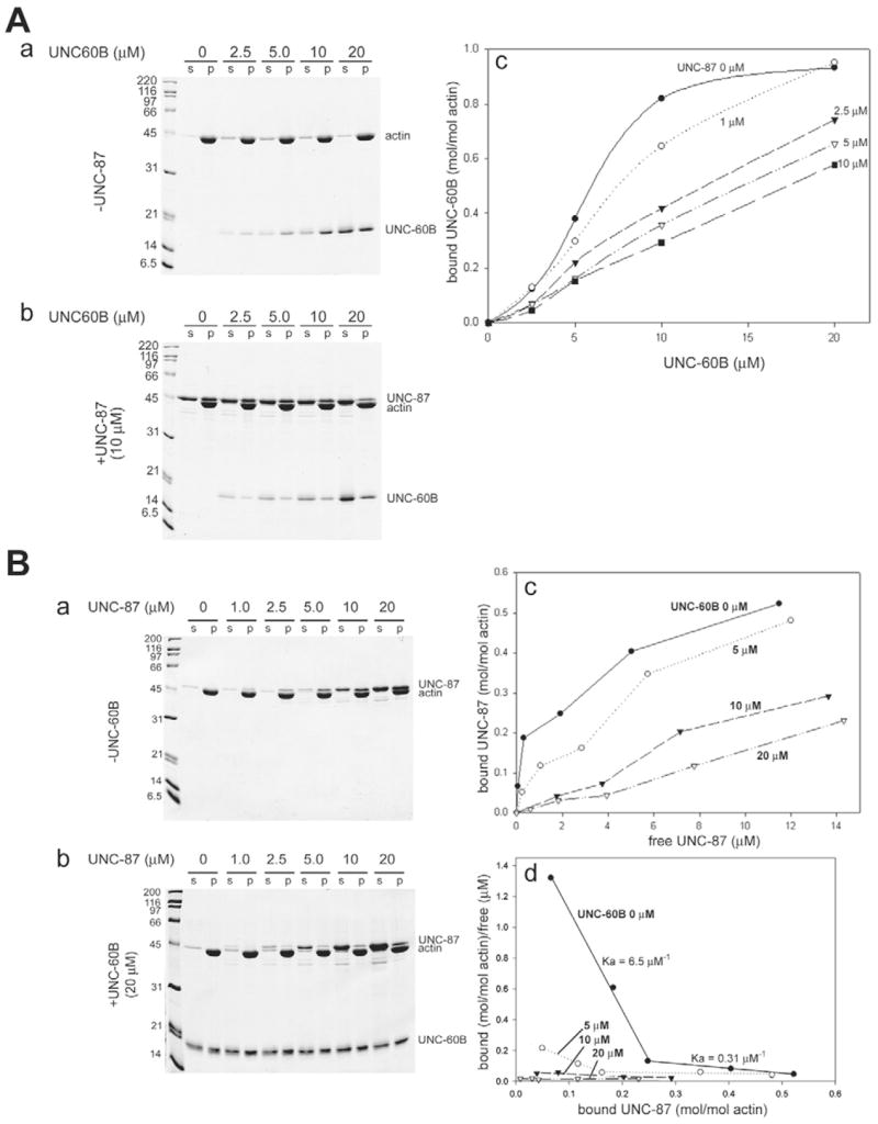
UNC-87 or UNC-60B interacts with Ce-actin in a mutually exclusive manner. (A) 10 μM Ce-actin was pre-incubated with 0–10 μM UNC-87 for 30 minutes, and then, various concentrations of UNC-60B (0–20 μM) were added to the mixtures. After 30 minutes, the samples were centrifuged at 285,000 g for 20 minutes, and the supernatant (s) and pellets (p) were analyzed by SDS-PAGE (12% acrylamide gel). Representative gels from experiments in the absence (a) or in the presence of 10 μM UNC-87 (b) are shown. Quantitative analysis of the results by densitometry is shown in c, in which the amounts of actin-bound UNC-60B (mol/mol actin) were plotted as a function of total UNC-60B concentrations (μM). (B) 10 μM Ce-actin was pre-incubated with 0–20 μM UNC-60B for 30 minutes, and then, various concentrations of UNC-87 (0–20 μM) were added to the mixtures. After 30 minutes, the samples were centrifuged at 285,000 g for 20 minutes, and the supernatant (s) and pellets (p) were analyzed by SDS-PAGE. Representative gels of experiments in the absence (a) and in the presence of 20 μM UNC-60B (b) are shown. Quantitative analysis of the results by densitometry is shown in c, in which the amounts of actin-bound UNC-87 (mol/mol actin) were plotted as a function of free UNC-87 concentrations (μM). (d) The quantitative results were further analyzed using a Scatchard plot.
