
Figure 5. Inoculation with macrophage inflammatory protein-3α plasmid (pMIP-3α) 3 days ahead of Gag plasmid (pGag) immunization results in improved interferon-γ (IFN-γ) expression in specific T cells.
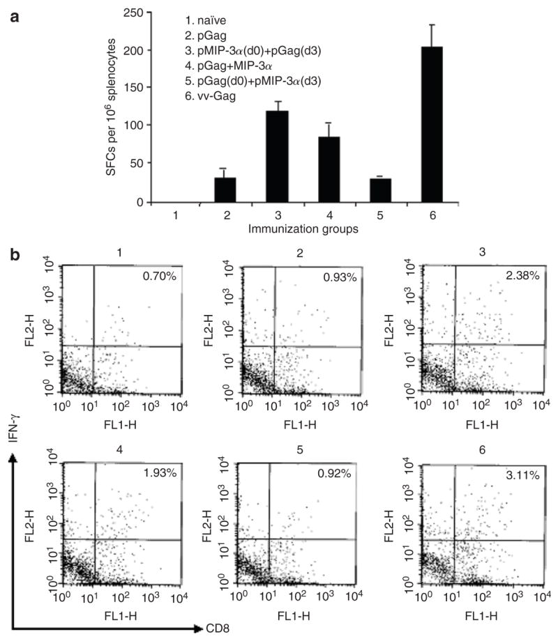
IFN-γ expression in Gag-specific T cells determined by (a) enzyme-linked immunosorbent spot (ELISPOT) and (b) intracellular staining. Mice were intramuscularly immunized twice, at weeks 0 and 2, with 50 μg of pGag alone or in conjunction with 50 μg of pMIP-3α, either 3 days before, simultaneous with, or 3 days after pGag vaccination. Animals were killed 2 weeks after the second immunization, and the splenocytes were harvested. Splenocytes were cultured for 30 hours in the absence or presence of 2 μg/ml of p7g peptide, and cells producing IFN-γ were detected using standard ELISPOT assay. For intracellular IFN-γ staining, two-color analysis on CD8 and IFN-γ was performed on splenocytes that had been previously stimulated at 5 × 10⁶ cells/ml for 30 hours with 2 μg/ml of p7g peptide. vv-Gag, vaccinia virus expressing Gag; SFC, spot-forming cell.