
Fig. 1.
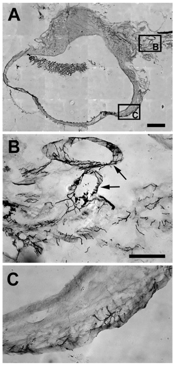
Photomicrographs of parts of a section immunostained with vesicular monoamine transporter (VMAT). A: Low-power view of area where photomicrographs in B and C were taken. B: at the entrance to the cyst, neurites are very dense and associated mainly with blood vessels (arrows) as they enter the wall of the cyst in the hilus region. C: further into the cyst, these neurites become less dense but also extend into the myometrium and epithelial lining. Note that this pattern was the same for neurites stained with other markers and for the uterine horn. Calibration bar: 500 μm for A; 100 μm for B and C.