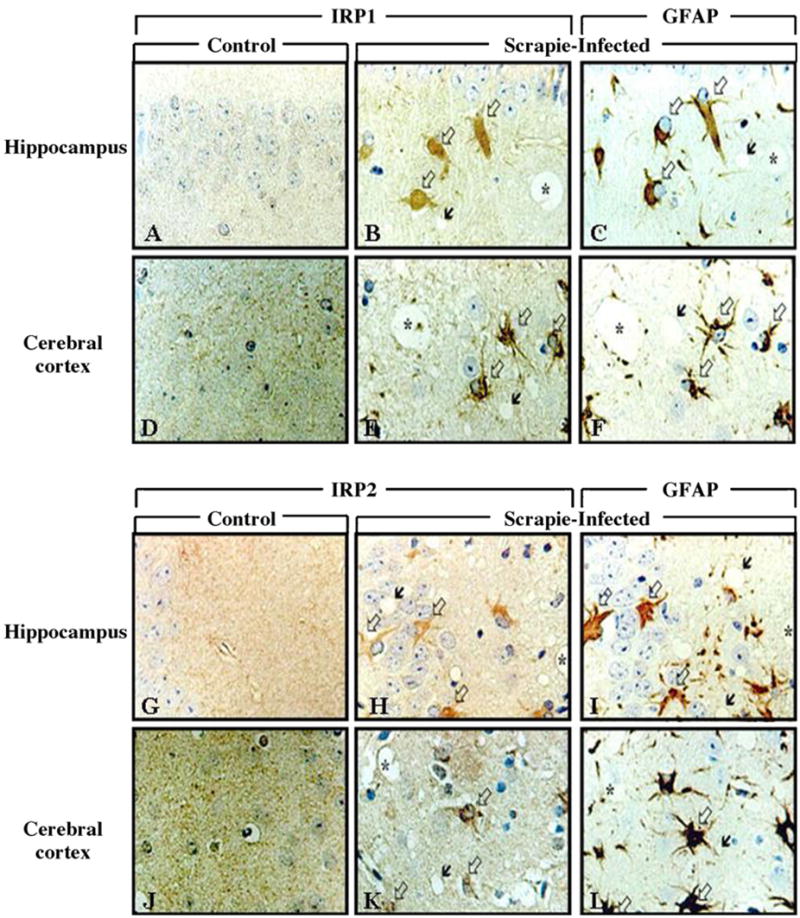
Fig. 2.

Immunohistochemical localization of IRP1 and IRP2 in the brains of control and scrapie-infected mice. Intense immunolabeling of IRP1 and IRP2 appear in the hippocampus and cerebral cortex of scrapie-infected mice. Black arrows: vacuoles, white arrows: immunolabeled cells with IRP1 (B and E) and IRP2 (H and K). In (C, F, I and L) white arrows indicate immunolabeling of GFAP and black arrows indicate vacuoles. In sequential sections, immunoreactivity of IRP1 and IRP2 is colocalized with GFAP-positive reactive astrocytes. Sequential sections are B and C, E and F, H and I, K and L. Asterisk (*) indicates landmark blood vessels in the adjacent tissue section.
