
Fig. 1.
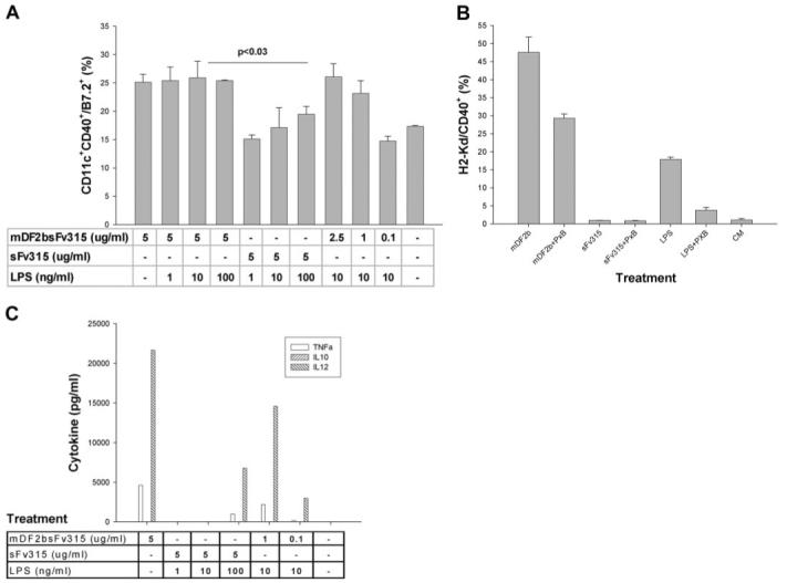
MDF2β activates APCs. (A) Maturation of XS52 cells was judged by up-regulation of CD11c+/CD40+/B7.2 cells after overnight incubation with indicated amounts (μg/ml) of mDF2β fusion protein (mDF2bsFv315). Control cells were treated with 5 μg/ml sFv315 in the presence or absence of indicated amounts of LPS (ng/ml). Comparable results were reproduced at least five times, and the mean of the representative triplicate experiment is shown ± sem. P < 0.03 is for comparison between treatments with mDF2β and sFv315. (B) Surface expression of H2Kd and CD40 on overnight-treated macrophage cell RAW264.7. Cells were treated with mDF2β-sFv315 (mDF2b; 5 μg/ml), sFv315 (5 μg/ml), or LPS (10 ng/ml) in the presence or absence of polymixin B (PxB; 5 μg/ml). Shown are representative data from four independent experiments with similar results ± sem. CM, Conditioned media. (C) ELISA results of cytokine production (pg/ml) in culture media of XS52 cells overnight treated with 5 μg/ml proteins or 10 ng/ml LPS in the presence or absence of 5 μg/ml polymixin B. The mean of the single representative triplicate experiment is shown.