
Fig. 4.
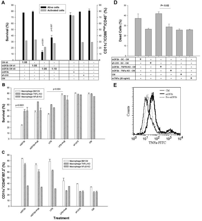
mDF2β induces death of APCs via activation of signaling cascades and secretion of cytotoxic factors. (A) Conditioned media from XS52 cells treated for 3 days with mDF2β (mDF2b CM d3) but not sFv315 are cytotoxic for naïve XS52 cells. In contrast, overnight conditioned media from overnight, mDF2β-treated XS52 cells are not toxic (mDF2b CM d1). Conditioned media are used at 1:5 and 1:10 dilutions. Cell survival (left y-axis) and activation (right y-axis) were measured as in Figure 3. The data were reproduced three times in triplicate experiments. (B and C) Experiments with macrophage cells with nonfunctional NF-κB (NFκB KO) and TNF-α (TNFα KO). mDF2β cannot kill (B) or activate (C) when cells do not express NF-κB. Moreover, cytotoxicity of mDF2β (B) is significantly abrogated when cells do not express TNF-α. The ability to be activated with mDf2β is not affected (C). Used, immortalized macrophage cells from B6/129 mice with KO p50 NF-κB or TNF-α genes, respectively. Controls were immortalized macrophage cells from B6/129 mice. Cell were treated for 3 days and stained in parallel to assess cell survival (B) or cell activation (C), as described in Figure 3. (D) Survival of naïve DCs incubated overnight with conditioned media (at dilutions 1:10) from wt DCs (mDF2b-DC-CM) or with nonfunctional genes TNFR1 (mDF2b-TNFR1-KO-CM) and TNF-α (mDF2b-TNFα-KO-CM) treated with 5 μg/ml mDF2β for 3 days. Control DCs were treated with 20 ng/ml TNF-α, or conditioned media were from sFv315-treated wt DCs (sFv315-DC-CM). Data were compared with effects of CM from XS52 cells treated with mDF2β (mDF2β-XS52-CM). (E) mDF2β (solid line) but not mproDF2β (dotted line) induces expression of cell-anchored TNF-α on the surface of the B6/129 macrophage. The black line is for untreated cells. Cells were stained with FITC-labeled anti-mouse TNF-α mAb after overnight incubation with proteins. No specific staining was detected with FITC-labeled, isotype-matched control antibody (data not shown). All results shown (A-E) were reproduced at least three times, and the mean of the representative triplicate experiment is shown ± sem. P values are for comparisons of between Day 1 and Day 3 conditioned media (A); wt and TNF-α KO or NF-κB KO macrophages treated with mDF2β or LPS (B and C); or between conditioned media of TNFR1 KO and TNF-α KO (D).