
Figure 3.
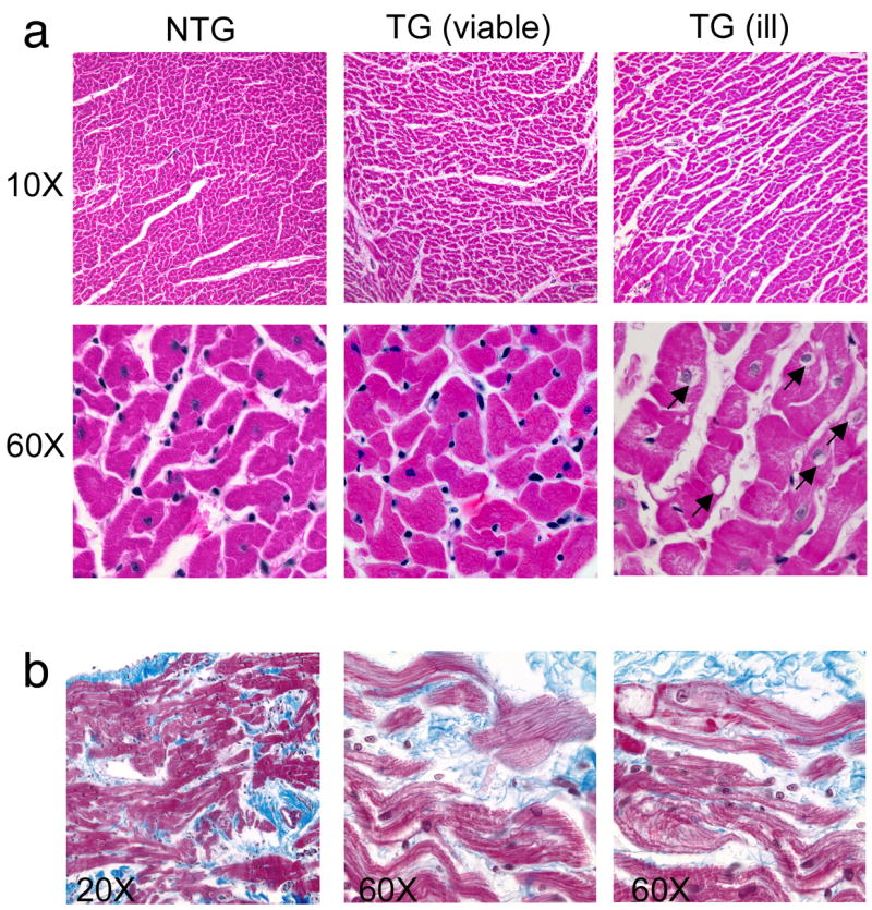
Cardiac histology of viable and ill transgenic lines. (a) All right ventricular sections were collected from 6 month old rabbits and stained with Masson’s trichrome. Ventricular myofibers from founder rabbits who were overtly ill exhibited a vacuolated appearance that was not observed in nontransgenic sections. Vacuoles are indicated by arrows. (b) Right ventricular sections taken from 6 month transgenic rabbits (viable line). Shown are areas of myocyte dropout, vacuolization and fibrosis that were not apparent in the nontransgenic hearts.