
Figure 4.
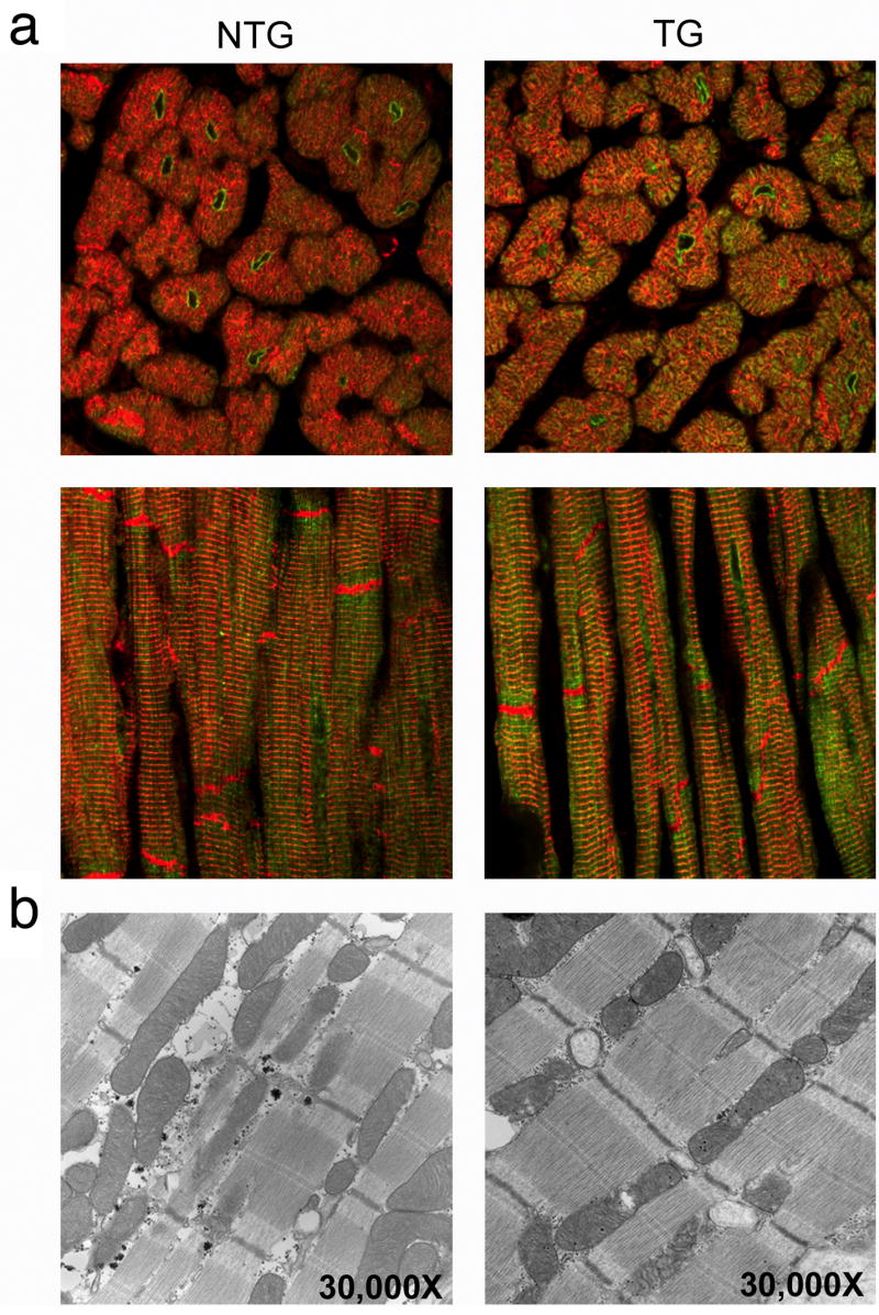
Phospholamban localization and ultrastructural analyses. (a) Whole heart sections from six month old rabbits were stained with phospholamban antibody (green) and counterstained with desmin antibody (red) to identify cardiac myofibers. No gender-specific differences were observed. Cross-sectional and longitudinal views of phospholamban staining show concentrated perinuclear accumulations in both the nontransgenic and transgenic sections. Magnification; 60X. (b) nontransgenic and transgenic hearts appeared to be identical at the ultrastructural level.