
Figure 1.
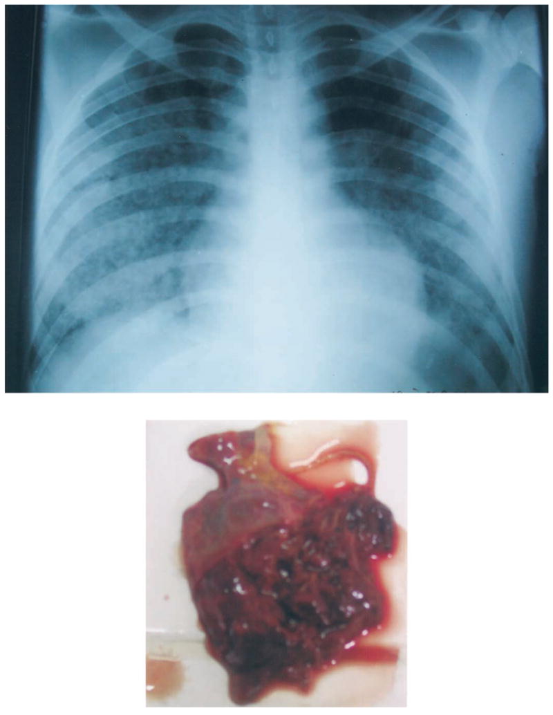
Chest radiograph (top) and gross appearance of right lower lung (bottom) from patient 4, a 19-year-old man who died from leptospiral pulmonary hemorrhage. The chest radiograph shows diffuse, patchy, alveolar infiltrates with a lower-lobe predominance. The right lower lung was extensively hemorrhagic and friable; the portion shown was obtained through the diaphragm through a peri-umbilical incision because only a partial necropsy was authorized.