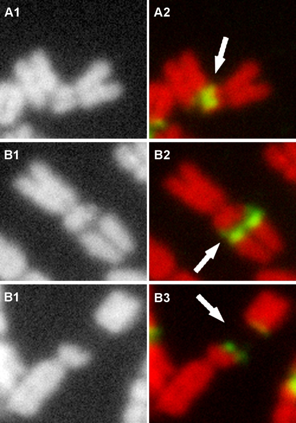
Figure 4. Different cytological appearances of lesions at the 45S rDNA fragile sites.
A: breakage or constriction occurs to a single chromatid within the 45S rDNA region. B: a gap forms within the rDNA between the two chromosome ends, but is still connected through one or a few thin DNA fibers (local despiralizations of the chromatid). C: A chromosome is broken and completely separated into two parts without any DNA hybridization signals detected within the gap. A1–C1: black layer; A2–C2: color image by merging red layers and green layers. Arrows indicate lesion sites.

