
Figure 5.
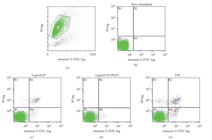
Cell apoptosis of different groups was detected by Annexin-V-propidium iodide staining. THP-1 cells were stimulated with 3 μg/mL of LP, 3 μg/mL of LP in combination with 25 μM PDTC, or 0.1 μg/mL LPS for 12 hours, stained with Annexin-V-FITC-PI and analyzed by FACS. Double negative staining represents living cells (c), positive staining for Annexin-V-FITC, and negative staining for PI represent the early apoptotic stage (e), and double-positive staining represents the late apoptotic stage (b).