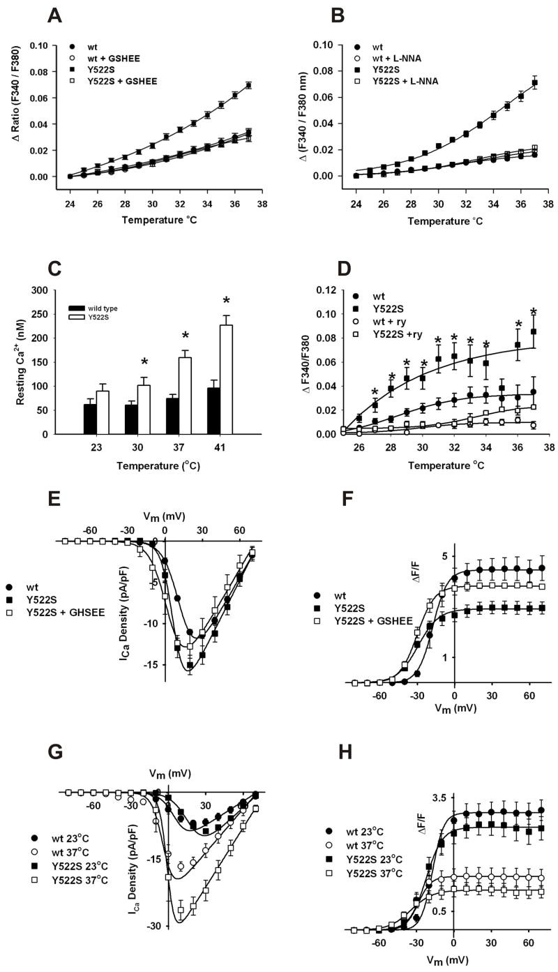
Figure 2. Temperature dependent increases cytosolic Ca2+ levels in RyR1Y522S/wt myotubes and solei.
A. Temperature dependent increases in cytosolic Ca2+ levels measured with fura-2. Myotubes loaded with fura-2AM were warmed to the indicated temperatures in the presence and absence of 5mM GSHEE. Values are mean ± SEM for 3 independent cultures for each group: RyR1wt/wt (●, n=27), RyR1wt/wt + GSHEE (○, n=31), RyR1Y522S/wt (■, n=29), RyR1Y522S/wt + GSHEE (□, n=32) (*p < 0.001, one-way ANOVA followed by Scheffe's comparison). B. Effect of L-NNA on temperature dependent increase in resting Ca2+. Myotubes loaded with fura-2AM were warmed to the indicated temperatures in the presence or absence of 50μM L-NNA. RyR1Y522S/wt (■, n= 27) RyR1Y522S/wt + L-NNA (□, n= 31), RyR1wt/wt (●, n= 28) and RyR1wt/wt + L-NNA (○, n= 33). *p<0.05, one way ANOVA followed by Scheffe’s comparison. C. Temperature dependent increases in cytosolic free Ca2+ concentration in RyR1Y522S/wt myotubes. Indo-1-loaded myotubes were warmed to the indicated temperatures and indo-1 ratios were calibrated as described in Methods. *p<0.05 compared to RyR1wt/wt at 23°C. D. Temperature dependent increases in cytosolic Ca2+ in solei fibers. Solei fibers of RyR1Y522S/wt mice were loaded with fura-2 and resting Ca2+ was measured in the presence or absence of ryanodine (20μM). RyR1Y522S/wt (■, n= 17), RyR1Y522S/wt + 20μM ryanodine (□, n=10), RyR1wt/wt (●, n= 12) and RyR1wt/wt + 20μM ryanodine (○, n= 4). *p<0.05, one-way ANOVA. E. Effects of GSHEE on the voltage dependence of L-type Ca2+ currents. Voltage dependence of average (± SEM) peak L-currents at room temperature in RyR1wt/wt myotubes (●), RyR1Y522S/wt myotubes (■), and RyR1Y522S/wt myotubes preincubated with 5 mM GSHEE (□). F. The effects of GSHEE on the voltage dependence of intracellular Ca2+ release. Ca2+ transients at room temperature were measured in RyR1wt/wt myotubes (●), RyR1Y522S/wt myotubes (■), and RyR1Y522S/wt myotubes preincubated with 5 mM GSHEE (□). G. Temperature dependence of L-type Ca2+ currents in RyR1wt/wt and RyR1Y522S/wt myotubes. Voltage dependence of average (± SEM) peak L-currents at 23°C (closed symbols) and 37°C (open symbols) in RyR1wt/wt myotubes (circles) and RyR1Y522S/wt (squares) myotubes. Each dataset was fit (smooth solid lines) using equations described previously (Chelu, 2006) in order to determine Gmax, VG1/2, kG, and Vrev at both 23°C (167 nS/nF, 7.8 mV, 8.6 mV, and 72.6 mV for RyR1wt/wt and 242 nS/nF, 17.3 mV, 7.1 mV, and 73.0 mV for RyR1Y522S/wt, respectively) and 37°C (309 nS/nF, −3.0 mV, 7.0 mV, and 72.5 mV for RyR1wt/wt and 442 nS/nF, −0.6 mV, 4.5 mV, and 78.2 mV for RyR1Y522S/wt, respectively). H. Temperature dependence of intracellular Ca2+ transients in RyR1wt/wt and RyR1Y522S/wt myotubes. Voltage dependence of average (± SEM) peak Ca2+ transient amplitude at 23°C (closed symbols) and 37°C (open symbols) in RyR1wt/wt myotubes (circles) and RyR1Y522S/wt (squares) myotubes Each dataset was fit (smooth solid lines) using equations described previously (Chelu, 2006) in order to determine Fmax, VF1/2, and kF at both 23°C (3.1, −16.8 mV, and 6.2 mV for RyR1wt/wt and 2.7, −23.1 mV, and 7.5 mV for RyR1Y522S/wt, respectively) and 37°C (1.4, −31.8 mV, and 5.7 mV for RyR1wt/wt and 1.0, −35.6 mV, and 8.2 mV for RyR1Y522S/wt, respectively for RyR1Y522S/wt, respectively). All data in this figure are shown as mean ± S.E.M.

