
Fig. 1.
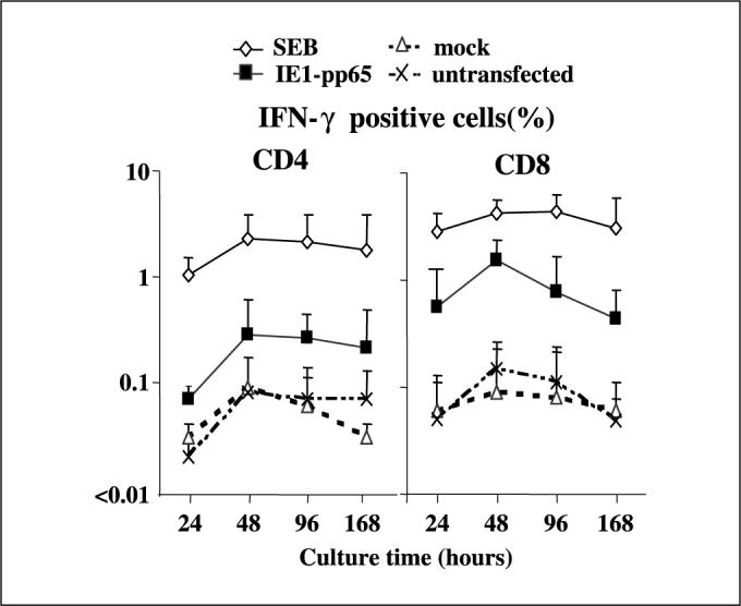
Maximum presentation of antigenic peptides processed from transfected protein by CD40B for both MHC class I and II occurs 48 hours after transfection. CD4 and CD8 T cells from four CMV-seropositive, HLA-A*0201–positive healthy donor PBMC were examined for IFN-γ production by ICC assay in response to IE1-pp65 fusion gene or mock transfected autologous CD40B. Both CD4 and CD8 Tcells showed maximum IFN-γ production after 48 hours culture with IE1-pp65 fusion gene transfectants at the 48-hour time point (0.3% and 1.6%, respectively). CD8 responses against transfectants were greater than those of CD4 Tcells but the transfected protein was processed with similar kinetics through both MHC class I and II pathways. SEB, Staphylococcus enterotoxin B (positive control); mock, CD40B cells transfected with vector alone; untransfected, CB40B cells alone (negative control). Points, mean; bars, SD.