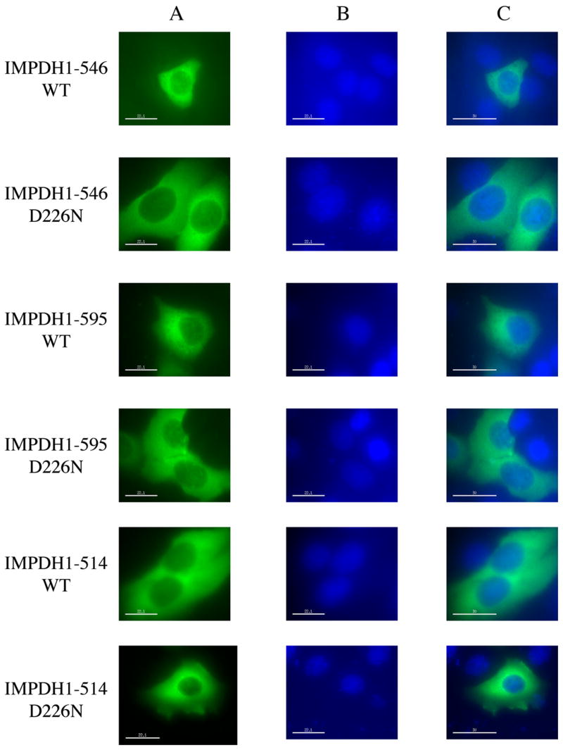
Figure 2.
Localization of the retinal IMPDH1 isoforms. HeLa cells transiently expressing IMPDH1-GFP were viewed under fluorescence microscopy. A) Distribution of various forms of IMPDH1 as revealed by the GFP fluorescence; B) Nucleic staining of the same cells by DAPI; C) Overlays of images in A and B.

