
Figure 3.
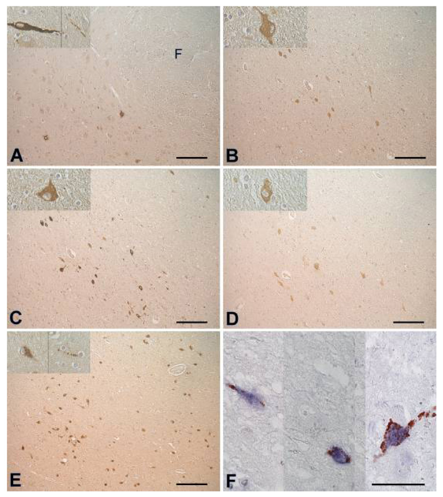
Adjacent sections from the lateral hypothalamus, stained with positive sera from 2 patients (A, B), 2 controls (C, D) and anti-hypocretin-1 (E). (F) 3 parts from sections double stained with anti-hypocretin-1 (blue) and serum of patient 1 (red). Note that no cell bodies are double stained. There are multiple bouton-like structures staining red, in close proximity of hypocretin cell bodies, suggesting nerve endings . Scale bars: (A–E): 200 µm, (F): 50 µm, (G): 1000 µm. Abbreviations: F = fornix, 3V = third ventricle. Figure reprinted with permission from 95