
Figure 3.
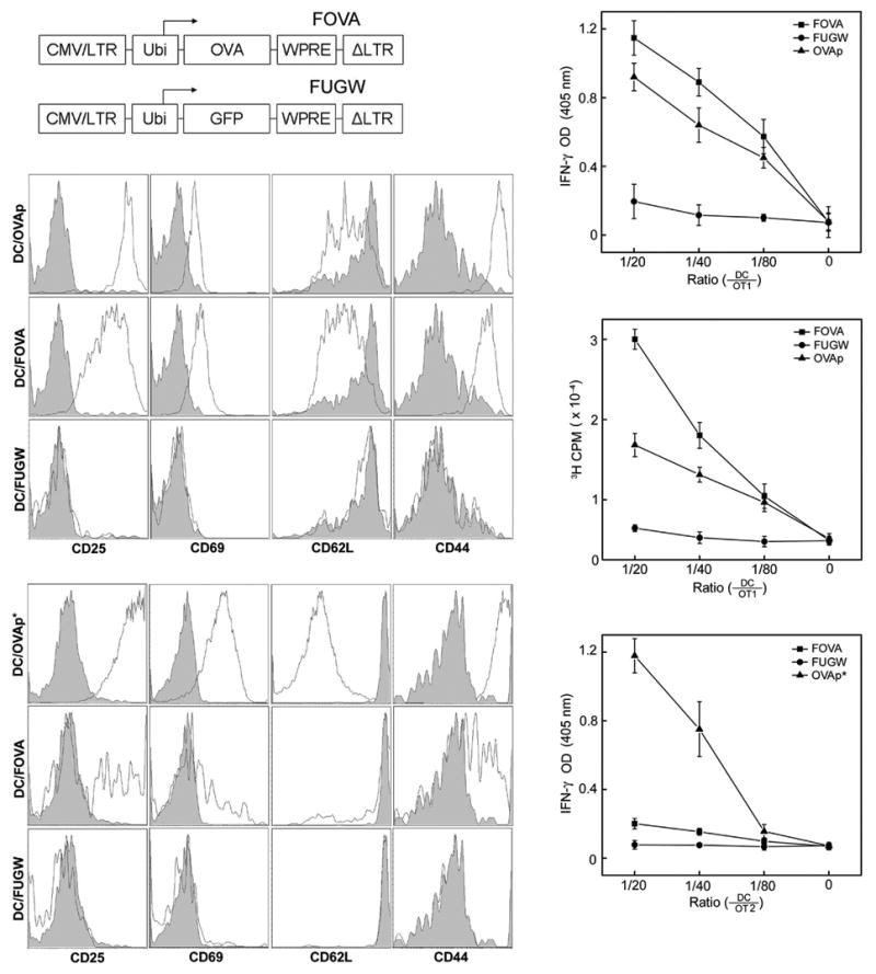
Mouse bone marrow-derived DCs (mBMDCs) transduced by a SVGmu enveloped lentivector encoding an OVA gene can stimulate OVA-specific CD8+ and CD4+ T cells in vitro. (a) A schematic representation of the lentivector encoding the OVA antigen (FOVA) or the lentivector encoding GFP (FUGW) as a control. LTR: long terminal repeat; Ubi: human ubiquitin-C promoter; WPRE: woodchuck hepatitis virus posttranscriptional regulatory element. (b-f) OVA-specific, CD8+ OT1 T cells and CD4+ OT2 T cells were harvested from the spleens of OT1 TCR- or OT2 TCR-transgenic mice (The Jackson Laboratory) and were cocultured with FOVA/SVGmu-transduced mBMDCs (DC/FOVA) in vitro for 3 days. Non-transduced BMDC pulsed with either OVAp peptide (SIINFEKL) (DC/OVAp), recognized by OT1 T cells, or OVAp* peptide (ISQAVHAAHAEINEAGR) (DC/OVAp*), recognized by OT2 T cells, were included as positive controls. mBMDCs transduced with FUGW/SVGmu (DC/FUGW) were included as a negative control. (b) Patterns of surface activation markers of OT1 T cells cocultured with various mBMDCs were assessed by antibody staining for CD25, CD69, CD62L, and CD44. Shaded area: naïve OT1 T cells harvested from transgenic animals; solid line: OT1 T cells cocultured with the indicated mBMDCs. (c) OT1 T cells were mixed with various dilutions of mBMDCs transduced with FOVA/SVGmu (■), FUGW/SVGmu (●), or pulsed with OVAp peptide (▲) and cultured for 3 days. Secretion of IFN-γ was measured by ELISA. (d) The proliferative responses of treated OT1 T cells from (c) were measured by a [3H] thymidine incorporation assay. (e) Similar to (b) except for the analysis of activated CD4+ OT2 T cells. Shaded area, naïve OT2 T cells harvested from transgenic animals; solid line, OT2 T cells cocultured with BMDCs. (f) Similar to (c) except for the responding cells being CD4+ OT2 T cells.